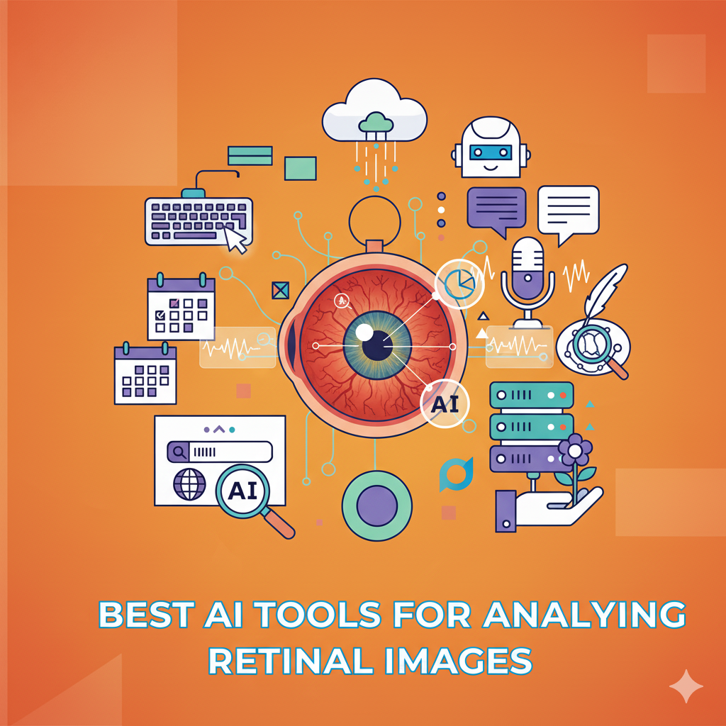
When we talk about healthcare and artificial intelligence, one of the most fascinating applications is in retinal image analysis. Our eyes can reveal a lot more than just vision problems retinal scans can help detect early signs of diabetes, hypertension, glaucoma, age-related macular degeneration (AMD), and even cardiovascular disease.
But analyzing retinal images manually takes expertise, time, and consistency. This is where AI-powered retinal image analysis tools are changing the game. They can process large volumes of scans quickly, highlight potential abnormalities, and support ophthalmologists in making faster, more accurate diagnoses.
In this article, let’s explore the best AI tools for retinal image analysis, their features, and what makes them stand out in future.
Why AI in Retinal Imaging Matters
Retinal imaging is critical because the eye is one of the few places where blood vessels can be observed directly without invasive procedures. Conditions like diabetic retinopathy and glaucoma often go undetected until it’s too late, which makes early diagnosis essential.
AI tools bring several advantages here:
- Early Detection – Identifies subtle changes invisible to the human eye.
- Scalability – Can handle thousands of scans efficiently.
- Accuracy – Reduces diagnostic errors and variability between doctors.
- Accessibility – Helps extend eye care to rural or resource-limited areas where specialists are scarce.
The Best AI Tools for Analyzing Retinal Images
1. Google’s ARDA (Automated Retinal Disease Assessment)
- What it does: ARDA is designed to detect diabetic retinopathy and other retinal conditions from fundus photographs.
- Key Features:
- FDA-approved for diabetic retinopathy screening.
- Provides automated, accurate results.
- Used in real-world clinical settings globally.
- Best For: Large-scale screenings, especially in regions with limited specialists.
2. Eyenuk EyeArt
- What it does: EyeArt is one of the most widely adopted AI-based diabetic retinopathy screening tools.
- Key Features:
- FDA-cleared and CE-marked.
- Fully autonomous delivers results in minutes without requiring eye care specialists.
- Cloud-based with integration support.
- Best For: Clinics and hospitals needing quick and scalable retinal disease screening.
3. Retmarker
- What it does: Retmarker specializes in diabetic retinopathy progression detection using AI.
- Key Features:
- Tracks disease progression over time.
- Helps ophthalmologists prioritize patients at higher risk.
- Integrates with existing retinal imaging systems.
- Best For: Long-term patient monitoring and clinical research.
4. IDx-DR (by Digital Diagnostics)
- What it does: The first FDA-cleared autonomous AI diagnostic system for diabetic retinopathy.
- Key Features:
- Provides immediate results without needing a human doctor to interpret images.
- Designed for use by primary care providers.
- HIPAA-compliant and highly secure.
- Best For: Primary care settings where immediate screening is required.
5. Zeiss VISUHEALTH AI Solutions
- What it does: Zeiss, known for its optical equipment, now integrates AI into its retinal imaging tools.
- Key Features:
- AI-assisted detection of glaucoma, AMD, and diabetic retinopathy.
- Seamless integration with Zeiss hardware and imaging devices.
- Trusted in ophthalmology clinics worldwide.
- Best For: Clinics already using Zeiss equipment looking for AI-powered upgrades.
6. Aira Matrix
- What it does: An AI platform focused on automated ophthalmic image analysis for clinical and research purposes.
- Key Features:
- Works with fundus and OCT (Optical Coherence Tomography) images.
- Cloud-based platform for multi-center usage.
- Provides detailed annotations for disease detection.
- Best For: Research institutions and hospitals requiring flexible AI image analysis.
7. DeepMind (now part of Google Health)
- What it does: DeepMind has developed AI algorithms that can detect 50+ eye diseases from retinal scans.
- Key Features:
- Published research in collaboration with Moorfields Eye Hospital, UK.
- Can match or exceed the diagnostic performance of ophthalmologists.
- Focuses on both screening and early intervention.
- Best For: Advanced research and large healthcare institutions.
How to Choose the Right AI Tool for Retinal Image Analysis
When selecting the best tool for your practice or institution, consider:
- Regulatory Approval – Is it FDA-approved or CE-marked for clinical use?
- Integration – Can it work with your existing imaging devices and workflows?
- Use Case – Do you need it for screening, diagnosis, or long-term monitoring?
- Scalability – Can it handle large patient volumes efficiently?
- Cost & Licensing – Subscription-based vs. one-time cost models.
The Future of AI in Retinal Imaging
AI in ophthalmology is still evolving. The next wave will focus on:
- Predictive analytics (forecasting disease progression).
- Telemedicine integration (remote screenings for rural populations).
- Multi-disease detection (using a single scan to check for multiple conditions).
- Personalized care plans (AI-driven recommendations for patient-specific treatments).
The future looks promising — AI won’t replace ophthalmologists but will act as a powerful assistant, making eye care more accurate and accessible.
Conclusion
The best AI tools for analyzing retinal images today include Google ARDA, EyeArt, Retmarker, IDx-DR, Zeiss AI solutions, Aira Matrix, and DeepMind. Each has its strengths, whether for large-scale screenings, long-term monitoring, or advanced research.
If you’re a healthcare provider or researcher, the key is to match the tool with your clinical workflow, budget, and patient needs. With AI, early detection and better eye care outcomes are not just possible — they’re becoming the new normal.


